Tampilkan postingan dengan label Skin Disease. Tampilkan semua postingan

Rabu, 19 September 2012

KELOID

What is a keloid?

Keloids can be considered to be "scars that don't know when to stop." A keloid, sometimes referred to as a keloid scar, is a tough heaped-up scar that rises quite abruptly above the rest of the skin. It usually has a smooth top and a pink or purple color. Keloids are irregularly shaped and tend to enlarge progressively. Unlike scars, keloids do not subside over time.

What is the difference between a keloid and a hypertrophic scar?

After the skin is injured, the healing process usually leaves a flat scar. Sometimes the scar is hypertrophic, or thickened, but confined to the margin of the wound. Hypertrophic scars tend to be redder and may subside by themselves (a process which can take one year or more). Treatment, such as injections of cortisone (steroids), can speed this process.
Keloids, by contrast, may start sometime after the injury and extend beyond the wound site. This tendency to migrate into surrounding areas that weren't injured originally distinguishes keloids from hypertrophic scars. Keloids typically appear following surgery or injury, but they can also appear spontaneously or as a result of some slight inflammation, such as an acne pimple on the chest (even one that wasn't scratched or otherwise irritated). Other minor injuries that can trigger keloids are burns and piercings.

What are keloid symptoms and signs?

Keloids are raised and look shiny and dome-shaped, ranging in color from pink to red. Some keloids become quite large and unsightly. Aside from causing potential cosmetic problems, these exuberant scars tend to be itchy, tender, or even painful to the touch.

Phototoxicity and photoallergy.

Phototoxicity and photoallergy.

Source

Department of Dermatology, University of California, San Francisco, USA.

Abstract

Photosensitivity may be phototoxic or photoallergic. Phototoxicity is much more common. There are 2 types of phototoxicity: photodynamic, which requires oxygen, and nonphotodynamic, which does not. Reactions induced by porphyrin molecules, coal tar derivatives, and many drugs are photodynamic. The reaction induced by psoralens, for the most part, is nonphotodynamic. Acute phototoxic reactions are characterized by erythema and edema followed by hyperpigmentation. Long-term ultraviolet phototoxicity results in chronic sun damage and skin cancer formation. Also, certain chemicals such as psoralen molecules and coal tar are photocarcinogenic. Phototoxic reactions to certain drugs produce unusual clinical patterns, that is lichenoid eruptions, dyschromia, photo-onycholysis, and pseudoporphyria. Photoallergy is an uncommon acquired altered reactivity dependent on an immediate antibody or a delayed cell mediated reaction. Solar urticaria is an example of the former, whereas photoallergy to exogenous chemicals is an example of the latter. Photoallergy to systemic drugs does occur but is difficult to characterize. The action spectrum for photoreactions to exogenous agents usually at least includes the ultraviolet A rays for both phototoxicity and photoallergy.

LIPOMA

Lipoma


What is a lipoma?

A lipoma is a growth of fat cells in a thin, fibrous capsule usually found just below the skin. Lipomas are found most often on the torso, neck, upper thighs, upper arms, and armpits, but they can occur almost anywhere in the body. One or more lipomas may be present at the same time. Lipomas are the most common noncancerous soft tissue growth.

What causes a lipoma?

The cause of lipomas is not completely understood, but the tendency to develop them is inherited. A minor injury may trigger the growth. Being overweight does not cause lipomas.

What are the symptoms of a lipoma?

Lipomas usually:
  • Are small [0.4 in. (1 cm) to 1.2 in. (3 cm)] and felt just under the skin.
  • Are movable and have a soft, rubbery consistency.
  • Do not cause pain.
  • Remain the same size over years or grow very slowly.
Often the most bothersome symptom is the location or increased size that makes the lipoma noticeable by others.

How are lipomas diagnosed?

A lipoma can usually be diagnosed by its appearance alone, but your health professional may want to remove it to make sure the growth is noncancerous.

How are lipomas treated?

Lipomas do not generally require treatment. Because lipomas are not cancerous growths and cannot become cancerous, they do not need to be removed. There is no known treatment to prevent lipomas or affect their growth.
A lipoma may be surgically removed if symptoms develop, such as if the lipoma:
  • Becomes painful or tender.
  • Becomes infected or inflamed repeatedly.
  • Drains foul-smelling discharge.
  • Interferes with movement or function.
  • Increases in size.
  • Becomes unsightly or bothersome.
Most lipomas can be removed in the doctor's office or outpatient surgery center. The doctor injects a local anesthetic around the lipoma, makes an incision in the skin, removes the growth, and closes the incision with stitches (sutures). If the lipoma is in an area of the body that cannot be easily reached through a simple incision in the skin, the lipoma may need to be removed in the operating room under general anesthesia.

Who is affected by lipomas?

Lipomas occur in all age groups but most often appear in middle age. Single lipomas occur with equal frequency in men and women. Multiple lipomas occur more frequently in men.

HIVES AND ANGIODEMA

Hives and Your Skin

Picture of Hives (Urticaria) Urticaria, also known as hives, is an outbreak of swollen, pale red bumps or plaques (wheals) on the skin that appear suddenly -- either as a result of the body's adverse reaction to certain allergens, or for unknown reasons.
Hives usually cause itching, but may also burn or sting. They can appear anywhere on the body, including the face, lips, tongue, throat, or ears. Hives vary in size (from a pencil eraser to a dinner plate), and may join together to form larger areas known as plaques. They can last for hours, or up to one day before fading.

What Causes Hives and Angioedema?

Hives and angioedema form when, in response to histamine, blood plasma leaks out of small blood vessels in the skin. Histamine is a chemical released from specialized cells along the skin's blood vessels.
Allergic reactions, chemicals in certain foods, insect stings, sunlight exposure, or medications can all cause histamine release. It's often impossible to find out exactly why hives have formed.
There are several different types of hives, including:
Acute urticaria: Hives lasting less than six weeks. The most common causes are certain foods, medications, or infections. Insect bites and internal disease may also be responsible.
The most common foods that cause hives are nuts, chocolate, fish, tomatoes, eggs, fresh berries, and milk. Fresh foods cause hives more often than cooked foods. Certain food additives and preservatives may also be to blame.
Drugs that can cause hives and angioedema include aspirin and other nonsteroidal anti-inflammatory medications such as ibuprofen, high blood pressure drugs (ACE inhibitors), or painkillers such as codeine.
Chronic urticaria and angioedema: Hives lasting more than six weeks. The cause of this type of hives is usually more difficult to identify than those causing acute urticaria. For most people with chronic urticaria, the cause is impossible to determine.
Chronic urticaria and angioedema can affect other internal organs such as the lungs, muscles, and gastrointestinal tract. Symptoms include muscle soreness, shortness of breath, vomiting, and diarrhea.
Physical urticaria: Hives caused by direct physical stimulation of the skin -- for example, cold, heat, sun exposure, vibration, pressure, sweating, and exercise. The hives usually occur right where the skin was stimulated and rarely appear elsewhere. Most of the hives appear within one hour after exposure.
Dermatographism: This is a common form of physical urticaria where hives form after firmly stroking or scratching the skin. These hives can also occur along with other forms of urticaria.

Heat rash

Symptoms

Adults usually develop heat rash in folds of skin and wherever clothing causes friction. In infants, the rash is mainly found on the neck, shoulders and chest, but it can also occur in the armpits, elbow creases and groin.

There are three types of miliaria, which are classified according to where the sweat ducts are blocked. Signs and symptoms for each type vary.

Miliaria crystallina
The mildest form of heat rash affects the sweat ducts in the topmost layer of skin. Miliaria crystallina is marked by:

  • Clear, fluid-filled blisters and bumps (papules) that break easily

The blisters that occur with miliaria crystallina aren't itchy or painful. This type of heat rash usually clears on its own but can come back if hot, humid weather persists. And though it's common in newborns, adults can develop it, too.

Miliaria rubra
Occurring deeper in the outer layer of skin (epidermis), miliaria rubra is sometimes called prickly heat. Adults usually develop miliaria rubra after they're exposed to hot, humid weather or if they're confined to bed rest. Infants usually develop this type of heat rash between the first and third weeks of life. Signs and symptoms typically include:

  • Red bumps

  • Itchy or prickly feeling in the affected area

  • Little or no sweating in the affected areas (anhidrosis)

Occasionally, miliaria rubra vesicles become pustular and then are called miliaria pustulosa.

Miliaria profunda
A less common form of heat rash, miliaria profunda occurs mainly in adults who have had repeat bouts of miliaria rubra. It affects the dermis, a deeper layer of skin, and appears soon after exercise or any activity that causes sweating. Signs and symptoms may include:

  • Firm, flesh-colored lesions that resemble goose bumps

  • A lack of perspiration, which may lead to symptoms of heat exhaustion, such as dizziness, nausea and a rapid pulse

When to see a doctor
Heat rash usually heals on its own and doesn't require medical care. See your doctor if you or your child has symptoms that last longer than a few days, the rash seems to be getting worse, or there are signs of infection, such as:

  • Increased pain, swelling, redness or warmth around the affected area

  • Pus draining from the lesions

  • Swollen lymph nodes in the armpit, neck or groin

  • A fever or chills

SCABIES


Scabies

Sarcoptes scabiei

Scabies is an easily spread skin disease caused by a very small species of mite.

Causes, incidence, and risk factors

Scabies is found worldwide among people of all groups and ages. It is spread by direct contact with infected people, and less often by sharing clothing or bedding. Sometimes whole families are affected.
Outbreaks of scabies are more common in nursing homes, nursing facilities, and child care centers.
The mites that cause scabies burrow into the skin and deposit their eggs, forming a burrow that looks like a pencil mark. Eggs mature in 21 days. The itchy rash is an allergic response to the mite.
Scabies is spread by skin-to-skin contact with another person who has scabies.
Pets and animals cannot spread human scabies. It is also not very likely for scabies to be spread by:
  • A swimming pool
  • Contact with the towels, bedding, and clothing of someone who has scabies, unless the person has what is called "crusted scabies"

Symptoms

  • Itching, especially at night
  • Rashes, especially between the fingers
  • Sores (abrasions) on the skin from scratching and digging
  • Thin, pencil-mark lines on the skin
Mites may be more widespread on a baby's skin, causing pimples over the trunk, or small blisters over the palms and soles.
  • In young children, the head, neck, shoulders, palms, and soles are involved.
  • In older children and adults, the hands, wrists, genitals, and abdomen are involved.

Signs and tests

Examination of the skin shows signs of scabies. Tests include an examination under the microscope of skin scrapings taken from a burrow to look for the mites. A skin biopsy can also be done.

Treatment

Prescription medicated creams are commonly used to treat scabies infections. The most commonly used cream is permethrin 5%. Other creams include benzyl benzoate, sulfur in petrolatum, and crotamiton. Lindane is rarely used because of its side effects.
Creams are applied all over the body. The whole family or sexual partners of infected people should be treated, even if they do not have symptoms. Creams are applied as a one-time treatment or they may be repeated in 1 week.
Wash underwear, towels, and sleepwear in hot water. Vacuum the carpets and upholstered furniture.
For difficult cases, some health care providers may also prescribe medication taken by mouth to kill the scabies mites. Ivermectin is a pill that may be used.
Itching may continue for 2 weeks or more after treatment begins, but it will disappear if you follow your health care provider's treatment plan. You can reduce itching with cool soaks and calamine lotion. Your doctor may also recommend an oral antihistamine.

Expectations (prognosis)

Most cases of scabies can be cured without any long-term problems. A severe case with a lot of scaling or crusting may be a sign that the person has a disease such as HIV.

Complications

Intense scratching can cause a secondary skin infection, such as impetigo.

Calling your health care provider

Call your health care provider if:
  • You have symptoms of scabies
  • A person you have been in close contact with has been diagnosed with scabies

Prevention

Avoid contact with infected persons.

BODY LICE


Body lice

Lice - body; Pediculosis; Vagabond's disease

Body lice are tiny parasites (Pediculus humanus corporis) that spread through close contact with other people.
There are three types of lice:
This article focuses on body lice.

Causes, incidence, and risk factors

Lice feed on human blood and live in the seams and folds of clothing for up to 1 month. They lay their eggs and deposit waste matter on the skin and clothing.
You can catch body lice if you come in direct contact with someone who has lice, or with infected clothing, towels, or bedding.
Body lice are bigger than other types of lice.
You are more likely to get body lice if you have poor hygiene or live in close (overcrowded) conditions. Infestation is unlikely to last on anyone who bathes regularly, and who has at least weekly access to freshly laundered clothing and bedding. Placing clothes in a hot dryer helps kill any lice on clothing.
If the lice fall off of a person, they die within about 5 - 7 days at room temperature.

Symptoms

Lice causes severe itching. It is usually worse around your waist, under the arms, near your bra strap, or anywhere clothing is tighter and closer to the body.
You may have red bumps on the skin. The bumps may scab or become crusty after scratching.
Skin around the waist or groin may become thickened or change color if the lice has been there a long time.

Signs and tests

Your doctor can usually diagnose this condition by looking at your skin and clothing.
  • Full-grown lice are the size of a sesame seed, have six legs, and are tan to greyish-white.
  • Nits are lice eggs. They will most often be seen in the clothing of someone with lice, most commonly around the waist and in the armpits.
Persons with body lice should also be checked for head and pubic lice.

Treatment

Body lice mainly live in clothing. Destroy or carefully wash infected clothing in hot water (at least 130 degrees F), then machine dry using a hot cycle.
Your doctor may also recommend a prescription cream or wash containing permethrin, malathione, or benzyl alcohol.
However, hygiene and washing clothes, bedding, and towels are most important and are usually enough.

Expectations (prognosis)

With effective treatment, the lice can be completely destroyed.

Complications

Scratching can make your skin more likely to become infected. Rarely, lice may carry uncommon diseases, such as trench fever. Because body lice is contagious, other family members may want to be treated.

Calling your health care provider

Call your health care provider if you have lice in your clothing or persistent itchiness.

Prevention

Good personal hygiene and effective treatment of infected persons will prevent lice from spreading to others.

MALIGNANT MELANOMA

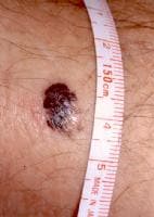
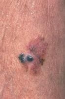
Malignant melanoma is a neoplasm of melanocytes or of the cells that develop from melanocytes. Although melanoma was once considered an uncommon disease, the annual incidence has increased dramatically over the over the past few decades, as have deaths from melanoma. (See the images of malignant melanoma below.) (See Etiology and Epidemiology.)
A 1.5-cm melanoma with characteristic asymmetry, iA 1.5-cm melanoma with characteristic asymmetry, irregular borders, and color variation. Malignant melanoma. Image courtesy of Hon Pak, MD.Malignant melanoma. Image courtesy of Hon Pak, MD. Also see Lentigo Maligna Melanoma, Oral Malignant Melanoma, and Head and Neck Mucosal Melanomas.

Growth

Melanomas have 2 growth phases, radial and vertical. During the radial growth phase, malignant cells grow in a radial fashion in the epidermis. With time, most melanomas progress to the vertical growth phase, in which the malignant cells invade the dermis and develop the ability to metastasize. (See Etiology and Workup.)
Clinically, lesions are classified as thin if they are 1 mm or less in depth; moderate if they are 1-4 mm in depth; and thick if they are greater than 4 mm in depth.

Histologic types of melanoma

There are 5 different forms, or histologic types, of melanoma:
  • Superficial spreading melanomas
  • Nodular melanomas
  • Lentigo maligna melanomas
  • Acral lentiginous melanomas
  • Mucosal lentiginous melanomas

Superficial spreading melanomas

Approximately 70% of cutaneous malignant melanomas are the superficial spreading melanoma (SSM) type and often arise from a pigmented dysplastic nevus. SSMs typically develop after a long-standing stable nevus changes; typical changes include ulceration, enlargement, or color changes. An SSM may be found on any body surface, especially the head, neck, and trunk of males and the lower extremities of females.

Nodular melanomas

Nodular melanomas (NMs) represent approximately 10-15% of melanomas and also are found commonly on all body surfaces, especially the trunk of males. These lesions are the most symmetrical and uniform of the melanomas and are dark brown or black. The radial growth phase may not be evident in NMs; however, if this phase is evident, it is short-lived, because the tumor advances rapidly to the vertical growth phase, thus making the NM a high-risk lesion. Approximately 5% of all NMs are amelanotic melanomas.

Lentigo maligna melanomas

Lentigo maligna melanomas (LMMs) also account for 10-15% of melanomas. They typically are found on sun-exposed areas (eg, hand, neck). LMMs may have areas of hypopigmentation and often are quite large. LMMs arise from a lentigo maligna precursor lesion. (See the image of lentigo maligna melanoma below.)
Lentigo maligna melanoma, right lower cheek. The cLentigo maligna melanoma, right lower cheek. The centrally located erythematous papule represents invasive melanoma with surrounding macular lentigo maligna (melanoma in situ). Image courtesy of Susan M. Swetter, MD.

Acral lentiginous melanomas

Acral lentiginous melanomas (ALMs) are the only melanomas that have an equal frequency among blacks and whites. They occur on the palms, soles, and subungual areas. Subungual melanomas often are mistaken for subungual hematomas (splinter hemorrhages). Like NM, ALM is extremely aggressive, with rapid progression from the radial to vertical growth phase.

Mucosal lentiginous melanomas

Mucosal lentiginous melanomas (MLMs) develop from the mucosal epithelium that lines the respiratory, gastrointestinal, and genitourinary tracts. These lesions account for approximately 3% of the melanomas diagnosed annually and may occur on any mucosal surface, including the conjunctiva, oral cavity, esophagus, vagina, female urethra, penis, and anus. Noncutaneous melanomas commonly are diagnosed in patients of advanced age. MLMs appear to have a more aggressive course than cutaneous melanomas, although this may be because they commonly are diagnosed at a later stage of disease than the more readily apparent cutaneous melanomas.

Sites other than the skin

The majority of melanomas are in the skin, but other sites include the eyes, mucosa, gastrointestinal tract, genitourinary tract, and leptomeninges. Metastatic melanoma with an unknown primary site may be found in lymph nodes only.

Staging

Clark staging is as follows:
  • level I - All tumor cells above basement membrane (in situ)
  • level II - Tumor extends into papillary dermis
  • level III - Tumor extends to interface between papillary and reticular dermis
  • level IV - Tumor extends between bundles of collagen of reticular dermis (extends into reticular dermis)
  • level V - Tumor invasion of subcutaneous tissue
Breslow classification (thickness) is as follows:
  • Less than or equal to 0.75 mm
  • 0.76-1.5 mm
  • 1.51-4 mm
  • Greater than or equal to 4 mm
The staging system for cutaneous melanoma was revised by the American Joint Committee on Cancer (AJCC) in early 2002.[1, 2]
AJCC groupings based on TNM classification are as follows:
  • Stage 0 - Tis, N0, M0
  • Stage IA - T1a, N0, M0
  • Stage IB - T1b, N0, M0; T2b, N0, M0
  • Stage IIA - T2b, N0, M0; T3a, N0, M0
  • Stage IIB - T3b, N0, M0; T4a, N0, M0
  • Stage IIC - T4b, N0, M0
  • Stage III - Any T, N 1-3, M0
  • Stage IIIA - pT1-4a, N1a, M0; pT1-4a, N2a, M0
  • Stage IIIB - pT1-4b, N1a, M0; pT1-4b, N2a, M0; pT1-4a, N1b, M0; pT1-4a, N2b, M0; pT1-4a/b, N2c, M0
  • Stage IIIC - pT1-4b, N1b, M0; pT1-4b, N2b, M0; any T, N3, M0
  • Stage IV - Any T, Any N, Any M
T classification (thickness) is as follows:
  • TX - Primary tumor cannot be assessed (shave biopsy, regressed primary)
  • Tis - Melanoma in situ
  • T1 - ≤1.0 mm (a: without ulceration, b: with ulceration)
  • T2 - 1.01-2.0 mm (a: without ulceration, b: with ulceration)
  • T3 - 2.01-4.0 mm (a: without ulceration, b: with ulceration)
  • T4 - < 4.0 mm (a: without ulceration, b: with ulceration)
N classification is as follows:
  • N1 - 1 lymph node; a: micrometastasis (clinically occult), b: macrometastasis (clinically apparent)
  • N2 - 2-3 lymph nodes; a: micrometastasis, b: macrometastasis, c: in transit met(s), satellite(s), without metastatic lymph nodes (N2a: 2-3 nodes positive for micrometastasis; N2b: 2-3 nodes positive for macrometastasis; N2c: In transit met(s) or satellite(s) without metastatic nodes)
  • N3 - 4 or more metastatic nodes or matted nodes or in-transit metastases or satellite(s) with metastatic node(s)
Note that micrometastases are diagnosed after elective or sentinel lymphadenectomy. Macrometastases are defined as clinically detectable nodal metastases confirmed by therapeutic lymphadenectomy or when nodal metastasis exhibits gross extracapsular extension.
M classification is as follows:
  • M1a - Distant skin, subcutaneous, or nodal metastases, normal lactate dehydrogenase (LDH) level
  • M1b - Lung metastases, normal LDH level
  • M1c - All other visceral metastases or any distant metastases with an elevated LDH level

BLEPHARITIS


Blepharitis

Eyelid inflammation
Blepharitis is inflammation of the eyelash follicles, along the edge of the eyelid.

Causes, incidence, and risk factors

Blepharitis is caused by an overgrowth of the bacteria that is normally found on the skin. It is usually due to seborrheic dermatitis or a bacterial infection. Both may occur at the same time.
The cause is overgrowth of the bacteria that is normally found on the skin.
Allergies and lice that affects the eyelashes may also cause blepharitis, although these causes are less common.
People who have blepharitis have too much oil being produced by the glands near the eyelid. This allows bacteria normally found on the skin to overgrow.
Blepharitis may be linked to repeated styes and chalazia. You are more likely to develop this condition if you have seborrheic dermatitis of the face or scalp, rosacea, lice, and allergies.

Symptoms

The eyelids appear red and irritated, with scales that stick to the base of the eyelashes. The eyelids may be:
  • Crusty
  • Reddened
  • Swollen
  • Itching
  • Burning
You may feel like you have sand or dust in your eye when you blink. Sometimes, the eyelashes may fall out.

Signs and tests

An examination of the eyelids during an eye examination is usually enough to diagnose blepharitis.

Treatment

Careful daily cleansing of the edges of the eyelids helps remove the skin oils that cause the bacteria to overgrow. Your health care provider might recommend using baby shampoo or special cleansers. Antibiotic ointments may also be helpful.

Expectations (prognosis)

The likely outcome is good with treatment. Continued attention to lid cleanliness may be required to prevent repeated problems. Continued treatment will typically make the eyes less red and more comfortable.

Complications

  • Styes
  • Chalazia
  • Injury to the eye tissue (corneal ulcer) from irritation
  • Inflammation of the surface of the eye (conjunctivitis)
  • Loss of eyelashes
  • Scarring of the eyelids

Calling your health care provider

Call for an appointment with your health care provider if symptoms worsen or do not improve after careful cleansing of the eyelids for several days.

Prevention

Cleaning eyelids carefully will help prevent blepharitis. If a specific skin condition is present, it should be treated.

HEAD LICE FACTS

Head lice facts

  • Head lice are parasites that are found on human heads. The word lice is plural for louse.
  • Head lice are spread by personal contact or the sharing of combs, brushes, caps, and other clothing.
  • Head lice are a common problem with preschool and schoolchildren.
  • Head lice cause a tickling feeling of something moving in the hair, itching, and sores on the head.
  • The affected individual, family members also infected, and the home all should be treated.
  • Remember: one head louse + one head louse = two head lice = the beginning of a head-lice infection.
  • Very young children should be evaluated by a health care professional before beginning medications.

What are head lice?

Head lice are parasites that can be found on the heads of people. Infection with head lice is called pediculosis. (The head louse, Pediculus humanus capitis, is different from the lice that cause body and pubic-hair infections.)

How common is head lice infestation?

Head lice infection is very common. It has been estimated that up to one in every 10 children in school acquires head lice at some time. In one study, the estimated annual cost of head lice infestations in the United States was nearly $1 billion dollars.

Who is at risk for getting head lice?

Anyone who comes in close contact with someone who already has head lice, or even their contaminated clothing and other belongings, is at risk for acquiring head lice. So it is easy to transmit head lice from one person to another. Preschool and elementary-school children (3-10 years of age) and their families are infected most often. Girls contract head lice more often than boys, and women contract more head lice than men. African-Americans rarely acquire head lice.

ACNE


Acne

Acne vulgaris; Cystic acne; Pimples; Zits
Last reviewed: November 22, 2011.
Acne is a skin condition that causes pimples or "zits." This includes whiteheads, blackheads, and red, inflammed patches of skin (such as cysts).

Causes, incidence, and risk factors

Acne occurs when tiny holes on the surface of the skin become clogged. These holes are called pores.
  • Each pore opens to a follicle. A follicle contains a hair and an oil gland. The oil released by the gland helps remove old skin cells and keeps your skin soft.
  • When glands produce too much oil, the pores can become blocked. Dirt, bacteria, and cells build up. The blockage is called a plug or comedone.
  • If the top of the plug is white, it is called a whitehead.
  • If the top of the plug is dark, it is called a blackhead.
  • If the plug breaks open, swelling and red bumps occur.
  • Acne that is deep in your skin can cause hard, painful cysts. This is called cystic acne.
Acne is most common in teenagers, but anyone can get acne, even babies. Three out of four teenagers have some acne. Hormonal changes may cause the skin to be more oily.
Acne tends to run in families. It may be triggered by:
  • Hormonal changes related to puberty, menstrual periods, pregnancy, birth control pills, or stress
  • Greasy or oily cosmetic and hair products
  • Certain drugs (such as steroids, testosterone, estrogen, and phenytoin)
  • High levels of humidity and sweating
Research does not show that chocolate, nuts, and greasy foods cause acne. However, diets high in refined sugars may be related to acne.

Symptoms

Acne commonly appears on the face and shoulders, but it may also occur on the trunk, arms, legs, and buttocks.

Signs and tests

Your doctor can diagnose acne by looking at your skin. Testing is usually not needed.

Treatment

SELF-CARE
Steps you can take to help your acne:
  • Clean your skin gently with a mild, nondrying soap (such as Dove, Neutrogena, Cetaphil, CeraVe, or Basics). Remove all dirt or make-up. Wash once or twice a day, including after exercising. However, avoid scrubbing or repeated skin washing.
  • Shampoo your hair daily, especially if it is oily. Comb or pull your hair back to keep the hair out of your face.
What NOT to do:
  • Try not to squeeze, scratch, pick, or rub the pimples. Although it might be tempting to do this, it can lead to skin infections and scarring.
  • Avoid wearing tight headbands, baseball caps, and other hats
  • Avoid touching your face with your hands or fingers.
  • Avoid greasy cosmetics or creams. Take off make-up at night. Look for water-based or "noncomedogenic" formulas. Noncomedogenic products have been tested and proven not to clog pores and cause acne.
If these steps do not clear up the blemishes, try over-the-counter acne medications. You apply these products directly to your skin.
  • They may contain benzoyl peroxide, sulfur, resorcinol, or salicylic acid.
  • They work by killing bacteria, drying up skin oils, or causing the top layer of your skin to peel.
  • They may cause redness or peeling of the skin.
A small amount of sun exposure may improve acne a little, but mostly it just hides the acne. However, too much exposure to sunlight or ultraviolet rays is not recommended because it increases the risk for skin cancer.
PRESCRIPTION MEDICINES
If pimples are still a problem, a health care provider can prescribe stronger medications and discuss other options with you.
Antibiotics may help some people with acne:
Creams or gels applied to the skin may be prescribed:
  • Retinoic acid cream or gel (tretinoin, Retin-A)
  • Prescription formulas of benzoyl peroxide, sulfur, resorcinol, or salicylic acid
  • Topical azelaic acid
For women whose acne is caused or made worse by hormones:
  • A pill called spironolactone may help
  • Birth control pills may help in some cases, though they may make acne worse
Minor procedures or treatments may also be helpful:
  • A laser procedure called photodynamic therapy
  • Your doctor may also suggest chemical skin peeling, removal of scars by dermabrasion, or removal, drainage, or injection of cysts with cortisone
People who have cystic acne and scarring may try a medicine called isotretinoin (Accutane). You will be watched closely when taking this medicine because of its side effects.
Pregnant women should NOT take Accutane, because it causes severe birth defects. Women taking Accutane must use two forms of birth control before starting the drug and enroll in the iPledge program. Your doctor will follow you on this drug and you will have regular blood tests.

Expectations (prognosis)

Acne usually goes away after the teenage years, but it may last into middle age. The condition often responds well to treatment after 6 - 8 weeks, but it may flare up from time to time.
Scarring may occur if severe acne is not treated. Some people, especially teenagers, can become very depressed if acne is not treated.

Calling your health care provider

Call your doctor or a dermatologist if:
  • Self-care measures and over-the-counter medicine have not helped after several months
  • Your acne is severe (for example, you have a lot of redness around the pimples or you have cysts)
  • Your acne is getting worse
  • You develop scars as your acne clears up
Call your baby's health care provider if your baby has acne that does not clear up on its own within 3 months.

DANDRUFF


Dandruff, also known as scurf or Pityriasis simplex capillitii, affects the scalp and causes flakes of skin to appear - it is a common condition. Our skin cells are forever renewing themselves. When the skin cells on our scalp are renewed the old ones are pushed to the surface and out of the scalp. For a person with dandruff the renewal is faster, meaning more dead skin is shed, making the dandruff more noticeable. Dandruff can also occur if the scalp is frequently exposed to extreme temperatures.

Dandruff can be chronical (long-term) or the result of certain triggers
. People with dandruff may also experience irritation and redness on the scalp.

Excessive flaking may be caused by an underlying illness or condition, such as psoriasis, a fungal infection (Malassezia), seborrheic dermatitis, or even head lice.

Some individuals with severe dandruff may have social or self-esteem problems. Therefore, treatment may be important for both physiological and psychological reasons.

The word dandruff comes from (most likely) dand (origin unknown) and E. Anglian (England) dialect huff, hurf, meaning "scab". This is probably linked to the Old Norse word hrufa, meaning "scab". The Old High German word hruf means "scurf".

According to MediLexicon's medical dictionary, dandruff is:

The presence, in varying amounts, of white or gray scales in the hair of the scalp, due to excessive or normal branny exfoliation of the epidermis.


A myth - some people think their dandruff is caused by their scalp being too dry. They try to deal with this by not washing their hair with shampoo, or wash it less often, believing that washing worsens the problem. This is a myth (not true). Dandruff differs from a dry scalp in that it usually gets better when you shampoo more frequently (with the right shampoos).


A significant number of people with dandruff find it improves as they get older.


It is estimated that about 50% of people in Western Europe and North America suffer from dandruff.


Dandruff is more common in men than in women, and in people with oily skin.


Some studies have suggested that diets that are too salty, sugary or spicy and accompanied by excessive alcohol may exacerbate dandruff.


Dandruff does not contribute to hair loss.

Minggu, 02 September 2012

Measles Treatment


Measles Treatment

There is no treatment that can kill the measles virus, so treatment focuses on supportive care, or the relief of symptoms. Supportive care can include intravenous fluids, medications to control fever or pain, and antibiotics to treat secondary infections from bacteria.


An Overview of Measles Treatment

There is currently no proven treatment that can kill the measles virus; therefore, measles treatment focuses on providing relief of measles symptoms as the body fights the virus. This is called supportive care.
Supportive Care for Treating Measles
Supportive care can include:
  • Intravenous (IV) fluids
  • Medications to control fever or pain
  • Antibiotics to treat secondary infections from bacteria
  • Good nursing care.
Vitamin A as Part of Measles Treatment
In developing countries, malnutrition, vitamin A deficiency, and severe measles are common. For these situations, the treatment of measles should include vitamin A medication for two days, starting as soon as a measles diagnosis is made. This treatment has been shown to decrease the risk of blindness and death.

What Is the Outcome Following Treatment?

The majority of people with measles recover, but measles complications can be dangerous. Six to twenty percent of the people who get the disease will get an ear infection, diarrhea, or even pneumonia. One out of 1,000 people with measles will develop inflammation of the brain, and about 1 out of 1,000 will die.

Chicken Pox

Chickenpox in Adults and Teenagers

Chickenpox causes a rash and can make you feel unwell. Symptoms tend to be worse in adults than children. Treatments can ease the symptoms until the illness goes. An antiviral drug may limit the severity of the illness if the drug is started within 24 hours of the rash first starting. Full recovery is usual. Serious complications are rare, but are more common in adults than children, and are more likely to occur in pregnant women and in people with a poor immune system.
chickenpox blisters
Chickenpox is an infection caused by the varicella-zoster virus. The immune system makes antibodies during the infection. These fight the virus and then provide lifelong immunity. Therefore, it is rare to have more than one bout of chickenpox in a lifetime.

Most people have chickenpox as a child. About 9 in 10 people have had it by the age of 15. It is uncommon for adults to have chickenpox.

Symptoms are usually more severe in adults than in children. Expect to have a few uncomfortable days.
  • Fever (temperature), aches and headache often start a day or so before a rash appears.
  • Rash. Spots appear in crops. The spots develop into small blisters and are itchy. They can be anywhere on the body and sometimes also in the mouth. Several crops may develop over several days. Some people are covered in spots; others have only a few.
  • Cough, sore throat, and feeling sick are common.
The fever and generally feeling unwell can last several days. The blisters gradually dry up and scab. They slowly fade over a week or so, but may take 2-3 weeks to go completely. A dry cough may persist for a while after all the other symptoms have gone.

Symptomatic treatments

Treatments that may ease symptoms whilst your immune system deals with the virus include the following:
  • Having plenty to drink to avoid dehydration.
  • Taking paracetamol or ibuprofen to ease fever, headaches, and aches and pains.
  • Calamine lotion put on the spots may ease itching.
  • Antihistamine tablets taken at bedtime may help you to sleep if itch is a problem at night. You can buy these at pharmacies, or get them on prescription.

Antiviral medication

Antiviral drugs such as aciclovir can limit the severity of chickenpox. These drugs do not kill the virus, but stop the virus from multiplying. Adults with chickenpox may be advised to take an antiviral drug - but only if the drug can be started within 24 hours of the rash first developing. If it is started after this time it is not likely to have much of an effect. So, if started on time, an antiviral drug may help to reduce the severity of the illness.

Antiviral medication is especially useful in situations where chickenpox can be more serious. For example, for people who have a poor immune system, newborn babies, and for pregnant women. (Note: antiviral medication is not normally advised for healthy children who develop chickenpox.)
  • The spots do not usually scar unless they are badly scratched.
  • In some cases, some spots become infected with bacteria. If this occurs, the surrounding skin becomes red and sore. Antibiotics may then be needed.
  • Inflammation of the lung (pneumonia) is a rare complication affecting about 1 in 100 adults with chickenpox.
  • Inflammation of the brain (encephalitis) is a very rare complication.
  • Vary rarely, other serious complications develop. For example: myocarditis (inflammation of heart muscle), glomerulonephritis (kidney inflammation), appendicitis, hepatitis (inflammation of the liver), pancreatitis (inflammation of the pancreas), Henoch-Schönlein purpura (a condition that affects kidneys), orchitis (inflammation of the testes), arthritis, and inflammation of various parts of the eye.
Therefore, although serious complications are rare, it is best to be vigilant. See a doctor if you develop any worrying symptoms that you are unsure about such as:
  • Breathing problems.
  • Weakness.
  • Drowsiness.
  • Convulsions.
  • Pains or headaches which become worse despite paracetamol or ibuprofen.
  • Being unable to take fluids due to a severe rash in the mouth.
  • A very severe rash, or a rash which bruises or bleeds into the skin ('haemorrhagic rash').
  • Becoming generally more and more unwell.
In general, complications are uncommon. However, some people have a higher risk of developing complications from chickenpox. Anyone in the following groups should see a doctor urgently if they have symptoms of chickenpox:
  • Pregnant women.
  • People with a poor immune system. For example, people with leukaemia, with HIV/AIDS, taking steroid medication or chemotherapy.
  • People with heart or lung disease.
  • People with severe skin conditions.
  • Very young babies - especially those less than a month old (neonates)..
A person with chickenpox is very infectious. The virus spreads in the air from person to person. For example, if you have not already had chickenpox, you stand a good chance of catching it if:
  • You are in the same room as someone with chickenpox for more than 15 minutes; or
  • You have any face-to-face contact with someone with chickenpox, such as a conversation.
It takes between 7 and 21 days (most commonly 10-14 days) to develop symptoms after catching the virus (the incubation period).
A person with chickenpox is infectious from two days before the rash first appears until all the spots have crusted over (commonly about five days after the onset of the illness). You can usually return to work after this time if you feel well enough. (A child with chickenpox should stay off school for five days from the onset of the rash.) Whilst you are infectious, keep away from people who have an increased chance of having a severe illness if they get chickenpox. These people are listed above under 'complications'.

Healthy people who have not had chickenpox may also want to avoid catching it. So, friends and family who have not had chickenpox may wish avoid you whilst you are infectious. However, most adults and many children have already had chickenpox, and so are immune.

Note: people with chickenpox should not travel by air until six days after the last spot appeared.

Yes, there is an effective vaccine that protects against the virus that causes chickenpox. It has become part of the routine childhood immunisation programme in certain countries such as the USA, Canada and Australia.

Currently, there are no plans to make immunisation against chickenpox routine for children in the UK. In the UK, the vaccine is offered by the NHS to certain special groups. For example, to healthcare workers who are not immune to chickenpox. Also, to people who are not immune to chickenpox and who are in close contact with people with a poor immune system - for example, brothers and sisters of children who are on chemotherapy.